When most vets think of alkaline phosphatase (ALP), what immediately comes to mind is the liver enzyme found on most chemistry panels. While that is indeed an important test for various hepatobiliary diseases, ALP is an enzyme with different isoforms found in many other cells throughout the body, including some that don’t usually contribute to serum levels. We can exploit the fact that ALP is found in a variety of cells to use that enzyme for a cytochemical reaction to help diagnose certain tumors.
We can exploit the fact that ALP is found in a variety of cells to use that enzyme for a cytochemical reaction to help diagnose certain tumors.
The best known use case for this is osteosarcoma (OSA). Fine needle aspirate cytology of lytic bone lesions can help differentiate obviously malignant lesions from benign proliferations such as a fracture or fungal osteomyelitis with reactive bone. However, discriminating an OSA from other types of sarcoma that can occur in bone is important for prognosis and formulating a diagnosis and treatment plan, but it can be difficult based on morphology alone.
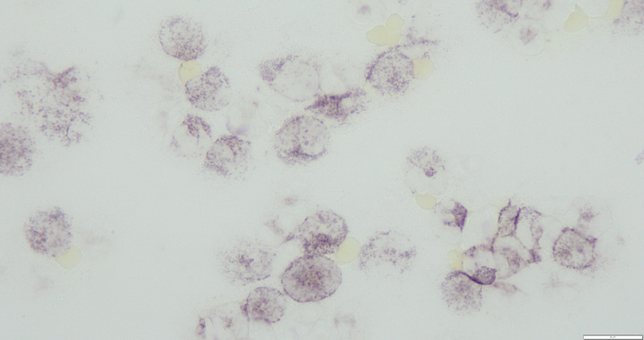
Since osteoblasts (both normal and malignant versions) express the bone isoform of ALP, we can use positive ALP staining in sarcoma cells to support a diagnosis of OSA. In one study evaluating the ability of ALP to diagnose OSA in vimentin-positive histopathology confirmed tumors, 33/33 OSAs were positive for a sensitivity of 100%, while specificity was 89% due to 1 of the 4 chondrosarcomas staining positive, along with one multilobular tumor of bone and one amelanotic melanoma. Another study found similar results, with a sensitivity of 88% and a specificity of 94% (non-OSA tumors included one each from a melanoma, gastrointestinal stromal tumor, histiocytic sarcoma, and anaplastic sarcoma).


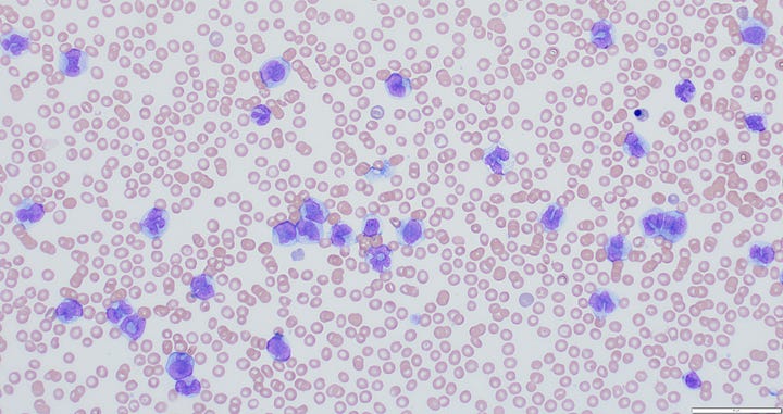
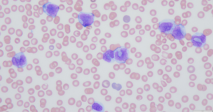
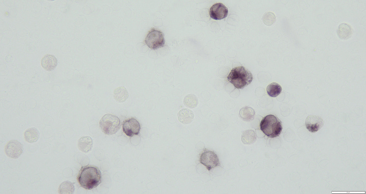
Another context that may be less familiar is using ALP to identify myeloid origin in poorly-differentiated leukemia cells. This can be useful since neoplastic cells from Acute Lymphoid Leukemia (ALL) and Acute Myeloid Leukemia (AML) may look very similar, but the prognosis differs dramatically, with AML having a grave course and typical survival times measured in days or weeks, while ALL is more likely to respond at least initially. A 2015 study of hematopoietic tumors found that ALP activity was present in 20/20 cases of AML (all monocytic or myelomonocytic) and absent in all cases of lymphoma and chronic lymphocytic leukemia (CLL). A minority of cases of T-cell ALL (31%) did show some ALP staining, although the authors commented: “the intensity of ALP staining was weak in all but one T-ALL dog and occurred in <20% of all neoplastic cells.” So the utility may improve if you account for how widespread and dark staining the ALP is among your tumor cells.


So, in conclusion, ALP staining on cytology can be a useful tool for several tumors, including osteosarcoma and acute myeloid leukemia. It is available in many university pathology labs, and increasingly several large commercial diagnostic companies. Many times your pathologist will recommend adding on this test (sometimes at an additional charge) based on their initial assessment. However, if you don’t see it mentioned and would like to run it, contact your lab to discuss the options!
References:
Barger A, Graca R, Bailey K, et al. Use of Alkaline Phosphatase Staining to Differentiate Canine Osteosarcoma from Other Vimentin-positive Tumors. Veterinary Pathology. 2005;42(2):161-165. doi:10.1354/vp.42-2-161
Ryseff JK, Bohn AA. Detection of alkaline phosphatase in canine cells previously stained with Wright-Giemsa and its utility in differentiating osteosarcoma from other mesenchymal tumors. Vet Clin Pathol. 2012 Sep;41(3):391-5. doi: 10.1111/j.1939-165X.2012.00445.x. Epub 2012 Jun 7. PMID: 22676437.
Stokol T, Schaefer DM, Shuman M, Belcher N, Dong L. Alkaline phosphatase is a useful cytochemical marker for the diagnosis of acute myelomonocytic and monocytic leukemia in the dog. Vet Clin Pathol. 2015 Mar;44(1):79-93. doi: 10.1111/vcp.12227. Epub 2014 Dec 26. PMID: 25546124.


