When Pathology Becomes Personal
Autopsies give us the facts but not the truth.
— Richard Selzer, MD
I. Anamnesis
1: a recalling to mind : REMINISCENCE
2: a preliminary case history of a medical or psychiatric patient
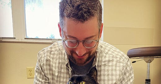
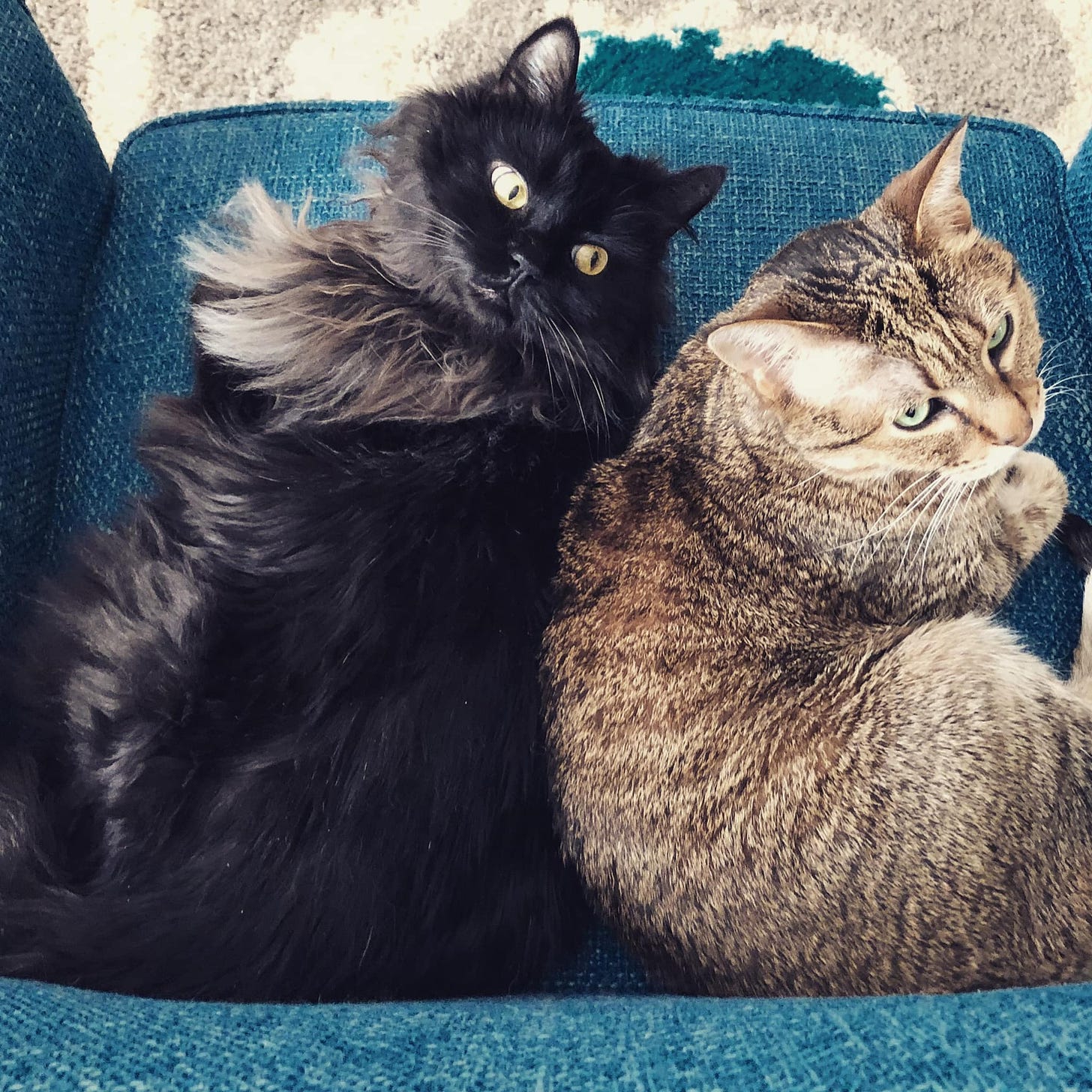
This is my first post since last August. One of the main reasons I got thrown off track with this site was the death of both my cats in 2022—Phoenix in March, followed by Ezra at the end of August, one day after my birthday. Their passing, along with travel and other events in my personal and professional life, distracted me and gave me a case of writer’s block. For a long time it was hard enough just to talk about their lives and deaths with family and friends, let alone discuss it with strangers. Weighed down by loss, the idea of writing anything, especially an irreverent science blog, felt pointless and trivial. Now that some time has passed and I’ve been able to process the grief, I feel ready to share some thoughts about what their lives and illnesses meant to me as a pet owner, a veterinarian, and a pathologist.
The stories of Phoenix and Ezra trace the arc of my educational and professional life. I adopted Phoenix while I was 21 and still a junior in college. He was several weeks old from a dairy farm on Lake Ontario in upstate New York. We originally thought this little ball of black fur was a female, but when it came time for his “spay” his vet realized he was actually a male, albeit with one of his testicles undescended (cryptorchid). Phoenix was one of the few constants in my nomadic life as I moved across the country to vet school in California, then to my internship in Virginia, residency in Alabama, and when my wife Lenore and I moved to Florida.
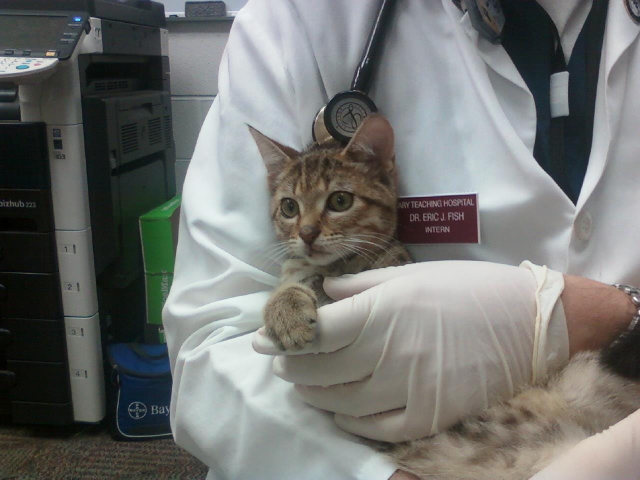
Ezra joined Phoenix during my internship. He was originally named “Tres” because they found him hiding under a Mazda 3 at twelve weeks old. The vet school didn’t have room for stray cats so they tried to cajole students and staff to adopt any they could. One community practice faculty member encouraged me to “test drive” Ezra by bringing him to rounds, and he was glued to my side and purred happily the whole time. I rationalized adopting him like many cat owners who have (incorrectly) thought, “I bet Phoenix is lonely and needs a friend, I should get a second cat.” And just like many owners have found out after bringing home the new feline competition, cat #1 most certainly did *not* want another one in the house! However, over time, his disdain for Ezra faded and the two became friends.
II. Treatment Failure (Phoenix)
From the time he was a kitten, Phoenix was a bit of a “lemon.” It started with him being cryptorchid. Then, when he was a little over a year, I had brought him into my study room at Western and he fell off a bookshelf he was climbing, leading to bilateral patellar luxation. A vet I worked for back home over summer breaks performed surgical repairs and noted his patellar groove was extremely shallow, a defect, one of many he’d develop over his life.
Phoenix was a great traveler, calmly riding in cars and planes with me without making a peep as I criss-crossed the country. One year coming home for winter break we were stuck at JFK airport overnight due to a blizzard. He was a trooper and peed on some pads I threw down in a handicap stall, but it was not a surprise when he developed straining in the litter box a few days later—a trip to the ER confirmed he had sterile feline lower urinary tract disease (FLUTD), which is often triggered by stress. From then on, he would have occasional flares of this that we would treat with diet, meds, and occasionally hospitalization.
When Phoenix was about 8 years old, he started off-and-on vomiting, as many cats do. We started changing his food to novel protein and hydrolyzed diets. They would help for a while, until they didn’t. Then began the endless lab tests and imaging. It eventually become clear that he had either lymphocytic IBD or small cell GI lymphoma, so we started treatment with prednisone, and that controlled his vomiting and weight for a while. Of course, as happens with some cats, the steroids led to diabetes, so we added insulin into the mix. Years of this went by: we would shuffle meds between prednisone, chlorambucil, and others, tweaking doses, adjusting insulin, as his clinical signs and weight fluctuated. Most of the time he was comfortable, ate well, had minimal vomiting or diarrhea, and seemed perfectly content.
In 2018 we started to lose control of his GI disease and diabetes. His appetite started to wane and he was losing weight. One day after bringing him to the internist for a recheck, I was getting back from the gym when Lenore called me upset: something was wrong with Phoenix. She had found him disoriented and collapsed with some bloody fluid around him. We rushed him to the ECC department at Auburn. He had experienced massive blood loss and was in shock. After some tests we discovered that his clotting times were abnormally prolonged, and he had likely not been absorbing Vitamin K as his GI disease and appetite worsened. This was a known, but rare, complication of his intestinal disease (it was barely documented in a few case reports from the 90s). Several transfusions and vitamin supplementation later, he made an amazing recovery and was discharged. We redoubled our efforts to control his GI disease and he slowly gained back weight and returned to his usual self.
Phoenix was stable and mercifully free of medical drama for several years until 2020. In the midst of the covid-19 pandemic, one day we found a hard lump on his left hock. Initial fine needle aspirate cytology identified a few spindle cells; as a clinical pathologist I was able to read between the lines on the report. We were almost certainly heading down the road to a cancer diagnosis. Follow-up biopsy revealed an aggressive Grade III sarcoma, most likely secondary to vaccination years ago. I was crushed, yet knew what we had to do—an amputation was the best way to save Phoenix’s life. He recovered from the surgery like a champ and along with some physical therapy, quickly became just as spritely and mobile as he was with four legs.
By 2022, Phoenix had survived a sarcoma and an alphabet soup of chronic internal medicine conditions. He took insulin injections like a champ. Chlorambucil gave his long fur a technicolor burst of gray ombre elegance. Things were in balance. Until they weren’t.
In February, Phoenix started inappropriately urinating around the house. This time, it was a dark orange-brown. We assumed that this was a flare-up of his previous FLUTD. Maybe this time he had a UTI secondary to his diabetes? With an upcoming trip, we decided it was best to take him to the vet.
I could tell something was wrong immediately from the internist’s tone of voice when they called with the initial results. Phoenix was in liver failure. His ALT, AST, ALP, and bilirubin were sky high. The orange tint to his urine were bilirubin byproducts, and despite being owned by two vets, we missed the telltale sign of jaundice. They were recommending additional tests, including fine needle aspiration of the liver. A few days later the pathologist’s report came back: Large Granular Lymphocyte (LGL) Lymphoma, a rare and aggressive form of cancer that has an almost invariably grave prognosis. I was devastated.
Besides the terrible news, the timing couldn’t be worse. I had an out of state work trip that week and Lenore and I had international travel booked for a few weeks later. We worked with the oncologists to try a chemotherapy protocol to buy us some amount of time, any. Despite our best efforts, Phoenix rapidly declined, losing weight and becoming frail.
Lenore called me when I was in Maine to let me know Phoenix looked terrible, and we decided it was time for humane euthanasia. We planned it for Friday afternoon when I returned from the trip. Unfortunately, Friday morning Phoenix began to slip into a coma. I was able to see him one last time and say goodbye over FaceTime from the seat of my plane before takeoff. He died about an hour before I landed in Tampa.
After 15 years of companionship and being a trooper with his various medical escapades, Phoenix was finally at peace.
III. Open Diagnosis (Ezra)
Compared to Phoenix, Ezra was healthy and low maintenance (at least in terms of health). He hated going to the vet and was terribly behaved at every step of the process, from trying to get him in the carrier to technicians and vets doing anything more invasive than pet him. We took him for annual wellness visits and his vaccines, but he never had any major issues.
By the age of 10, Ezra started to vomit just like his brother. Here we go again, I thought… Unlike Phoenix, his labwork was always boring and we did abdominal imaging from time to time that didn’t show anything concerning. That’s why when he started vomiting last August it wasn’t immediately cause for alarm. However, within a few days, the frequency and intensity was abnormal for him. Ezra seemed tense and uncomfortable in the abdomen, so we took him to a nearby specialty hospital for evaluation and treatment.
The first thing they noticed was he was running a high fever, in the 104-105F range. We all suspected an infection, perhaps he had swallowed a foreign object and it was causing an obstruction? Yet abdominal x-rays and ultrasound were negative for obstruction, free fluid, masses, enlarged organs, or other abnormalities. As usual, his bloodwork didn’t point in any specific direction.
Over the next few days, Ezra developed low blood pressure, further increasing our suspicion for an occult infection. We pulled a urine culture, started IV fluids, pain medications, and powerful anti-nausea drugs while we discussed expanded infectious disease testing and blood cultures. The vets also started multiple broad-spectrum IV antibiotics while we waited for the culture to come back (it was ultimately negative). With no response to antibiotics and his condition worsening, we started that ultimate veterinary Hail Mary treatment: steroids. Alas, like all of the other interventions, those did nothing to improve his fever, hypotension, or other clinical signs.
Because he was so stressed in the hospital—we could hear him hissing and growling for the nurses while we waited outside the ICU—we tried taking him home to see if his anxiety and pain would be better on an outpatient basis. However, he spent the whole time hiding and vomiting, and we brought him back for re-admission the next day.
We kept him in the hospital on my birthday, hoping I could enjoy the day and that he might pull through. The next morning, the doctors called us to say he was critically ill and we should start having discussions about quality of life.
As we sat with Ezra in the waiting room, I cradled him and ran through the bargaining stage of grief: Surely there was something we missed, some key fact that would point to why he was suddenly so ill, and potentially something we could treat. Maybe we missed a GI perforation after all! We rechecked abdominal imaging. It was still normal. Maybe we could find the source if we did exploratory abdominal surgery! At this point, Lenore gently helped me move past the denial stage into acceptance.
We euthanized Ezra that afternoon, and for the second time in a few months, I was crushed by the loss of a beloved pet. Besides the obvious grief, I was haunted by the possibility that we missed something. He didn’t have to die, I told myself. The hospital asked how we wanted to process the remains. Would we be interested in an autopsy? For the second time that year, I declined a post-mortem and elected cremation.
IV. Post-Mortem
The word “autopsy” first came into use in the 1600s, and is derived from the Greek roots “auto” for self, and “ops” for eye, meaning roughly “to see for one’s self.” This burgeoning practice of a diagnostician investigating the cause of death was a novel development from the late Renaissance when the primary purpose of such a post-mortem examination (often on the remains of prisoners, political dissidents, or even bodies exhumed by grave-robbers) was to teach normal anatomy to medical students and scientists.
Around the mid-twentieth century, approximately half of all deaths in a hospital setting were investigated by autopsy. Today, that rate has plummeted to less than 1 in 20. Such a dramatic decline is worrisome, because multiple studies have shown substantial disagreement with the antemortem clinical diagnosis and the final autopsy diagnosis, approximately 25%. This decline in post-mortems has also reduced student and clinician exposure to the art and science of opening the body, examining the organs and tissues, sifting through mortal questions that cannot be asked of the living.
As a veterinarian in practice and later a pathologist, I was often frustrated when clients would decline a necropsy (term of art for an autopsy on a non-human subject) after their pet died. In my experience, very few if any patients ever receive a necropsy. So many cases end without closure. What was the final diagnosis? Why did that treatment or surgery fail? Could anything have altered the course of the disease?
Despite all of that, I chose not to have either Phoenix or Ezra receive a necropsy. This surprised me as I always thought of myself as the type of person who needed to know the final answer, regardless of the outcome. However, in the moment, this felt like the right decision for each.
For Phoenix, the underlying diagnosis was clear and unambiguous: A final report would only confirm what had been seen through less invasive testing: cancer cells that had been seen on cytology infiltrating all of the major abdominal organs; hepatic insufficiency that caused his visible jaundice, spiking liver enzymes and bilirubin; evidence of hepatic encephalopathy from excessive ammonia in the blood that hopefully eased his final moments into a quiet sleep. The only remaining questions were hypotheticals that could never be answered: Was there something in his genes or previous medical conditions that predisposed to this rare form of lymphoma? Did his years of chlorambucil mutate a rogue cell that went dormant until those last weeks? What if we had caught the LGL lymphoma sooner? What if we tried a different chemotherapy regimen?
On the other hand with Ezra, we still don’t know why he became so ill and rapidly declined. Our working theory was sepsis, but we could never find a source, and he did not respond to antibiotics. I worried that he had ingested a foreign body that could have obstructed his GI, but serial radiographs and ultrasound did not support that possibility. Prior to deciding on euthanasia, we discussed exploratory surgery in case there was something lurking not detectable by blood analyzers or radiowaves, but in the end, that in itself carried a risk of morbidity and mortality, as well as the result of a negative explore. There is a strong possibility a necropsy could have given me a final diagnosis and closure. However, I was terrified that it would find an easily treatable problem we had simply missed or not considered, and having an avoidable death on my hands would have shattered me.
Finally, as Richard Selzer so eloquently stated at the top of this essay, an autopsy would simply provide a list of cold facts that may or may not shed any light on what was happening in their bodies. It would say nothing about their colorful lives or the people they touched. It would be mute about whether or not either Phoenix or Ezra’s outcomes would be any different if we knew more about the cellular pathology.
V. Afterword
As I reflect on the lives of both Phoenix and Ezra nearly a year later I am filled with joy for all of the time we were able to spend together. For all of the pain of their passing, there were so many more moments of absurdity and serendipity. I remember Phoenix climbing on top of the refrigerator to glare down on us despite his bad knees and how he used to steal food out of cabinets and the table like a ninja. I remember how Ezra loved our dogs and would snuggle in to spoon in under the covers at night.
I’m also grateful for all of the medical lessons I learned. It is so easy for a veterinarian to create distance between themselves and their patients and diseases, and a pathologist is even further removed from laying hands on animals or talking to owners. All too often I’ve diagnosed patients with an aggressive form of terminal cancer or an intractable infection and felt more awe at the terrible beauty of the microscopic lesion than considered the suffering of the animal connected to those cells or their family. In some sense, this dissociation is a necessary coping mechanism; as the covid-19 pandemic showed, the end result of being immersed in constant bad news and grief is compassion fatigue, burnout, or worse. Yet, experiencing personal losses like Ezra and Phoenix served as a reminder of the importance of what I do for a living, and it refreshed the well of empathy for the thousands of people and animals whom I indirectly change forever with a few sentences in a report.
It was also a humbling reminder of the limits of modern medicine and knowledge itself. Despite having access to nearly all of the latest diagnostics from human medicine, some diseases refuse easy diagnosis or classification. Even though we have made huge advances in veterinary oncology, cures remain elusive, and some types are completely refractory to treatment. Those of us in healthcare are forced to admit on a daily basis that we don’t know everything, we are simply doing our best, and all we can hope for is extending the time we have with the best quality possible.


