Dear Readers,
For this edition of “Five Minute Paper,” I will be walking through the first large(-ish) study of canine granular lymphoma. This rare form of cancer is quite aggressive and it may occur in both cats and dogs. In fact, this is ultimately what my cat Phoenix died from in 2022. Understanding details about this disease are important for vets to know how to treat it and counsel pet owners when it strikes.
The Study:
Yale AD, Crawford AL, Gramer I, Guillén A, Desmas I, Holmes EJ. Large granular lymphocyte lymphoma in 65 dogs (2005-2023). Vet Comp Oncol. 2023 Dec 29. E-pub ahead of print.
Abstract
Large granular lymphocyte lymphoma (LGLL) is a rare form of lymphoma in dogs. Limited information exists regarding presentation, treatment response, and outcome. The aim of this single-institute, retrospective study was to characterise clinical presentation, biologic behaviour, outcomes, and prognostic factors for dogs with LGLL. Cytologic review was also performed. Sixty-five dogs were included. The most common breed was the Labrador retriever (29.2%), and the most common presenting signs were lethargy (60.0%) and hyporexia (55.4%). The most common primary anatomic forms were hepatosplenic (32.8%) and gastrointestinal (20.7%). Twenty dogs (30.8%) had peripheral blood or bone marrow involvement. Thirty-two dogs were treated with maximum tolerated dose chemotherapy (MTDC) with a response documented in 74.1% of dogs. Dogs ≥7 years, and those with neutropenia or thrombocytopenia at diagnosis had the reduced likelihood of response to treatment. For dogs treated with MTDC median progression-free interval (PFI) was 17 days (range, 0-481), the median overall survival time (OST) 28 days (range, 3-421), and the 6-month and 1-year survival rates were 9.4% and 3.1%, respectively. On multivariable analysis, monocytosis and peripheral blood involvement were significantly associated with shorter PFI and OST. Long-term survival (≥100 days) was significantly associated with intermediate lymphocyte size on cytology. Dogs with LGLL have moderate response rates to chemotherapy but poor overall survival. Additional studies are needed to further evaluate prognostic factors and guide optimum treatment recommendations.
What is this study about?
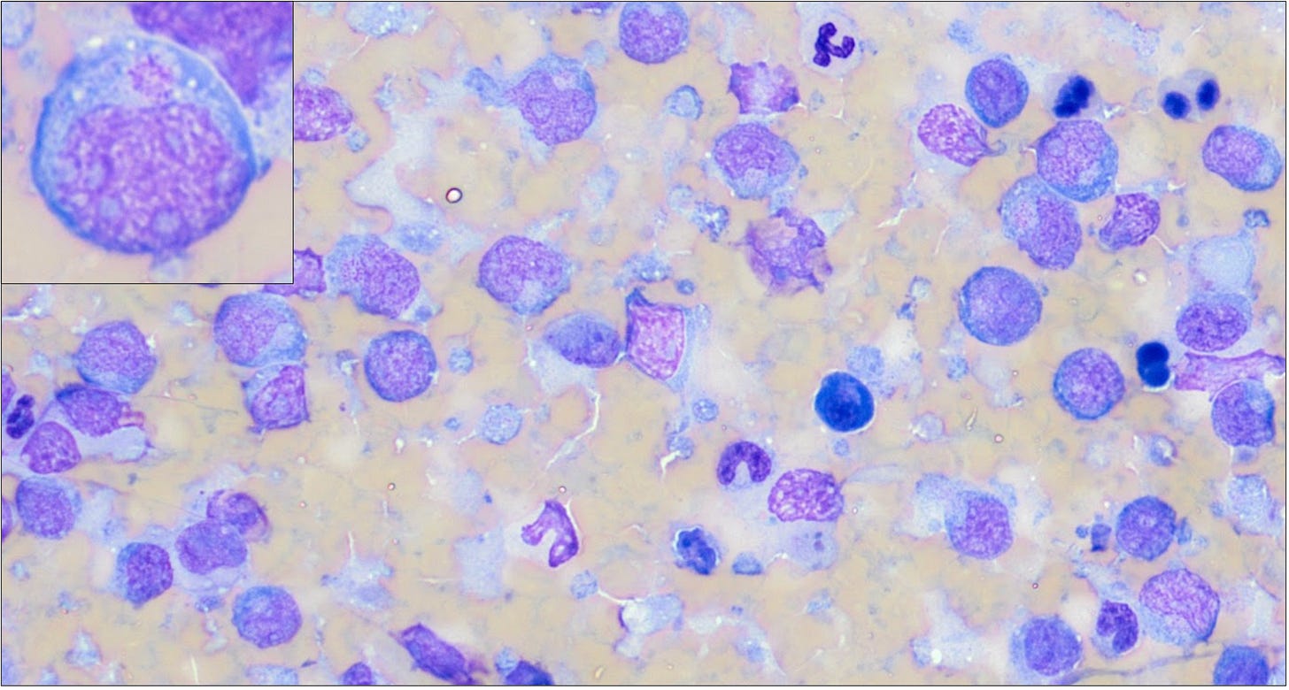
The study, based on cases from 2005 to 2023, examined large granular lymphocyte lymphoma (LGLL) in 65 dogs. The disease is named for the characteristic pink to magenta granules in the cancer cell cytoplasm; these are thought to contain perforin, granzyme, and other cell-killing enzymes produced by cytotoxic T cells and/or NK cells. The researchers aimed to characterize the clinical presentation, biologic behavior, outcomes, and prognostic factors of this rare form of lymphoma.
Who did the research?
This study was a collaboration between researchers in the (1) Department of Clinical Science and Services and (2) Department of Pathobiology and Population Sciences at the Royal Veterinary College in the UK.
Who paid for it? Any conflicts of interest?
The authors report no funding was used for this study and they declared no conflicts of interest.
What did they do?
This was a retrospective observational study. Records were searched for canine patients seen from 2005 to 2023. Inclusion criteria were a diagnosis of intermediate or large granular lymphoma by cytology and/or histopathology; dogs with a proliferation of only small cell granular lymphocytes or only blood/marrow involvement were excluded as this disease behaves differently, with prolonged survival. A board-certified veterinary clinical pathologist reviewed all available cytology slides. Treatment responses were based on cRECIST v1.0 or the VCOG response evaluation criteria for peripheral nodal lymphoma. Statistics included Kaplan-Meier survival analysis and other basic hypothesis testing using SPSS software.

What did they find?
Clinical characteristics
65 dogs included in final study population
The median age was 8.9 years (range: 1.3–14.8)
Labrador Retrievers were the most commonly affected breed (29.2%)
Other common breeds included: mixed breed (n=5 [7.7%]), border collie (n=4 [6.2%]), golden retriever (n=4 [6.2%]), and Staffordshire bull terrier (n=4 [6.2%])
Most dogs (61/65, 93.8%) presented sick in WHO substage ‘b’
Common clinical signs were lethargy (60%), decreased appetite (55.4%), vomiting (n=22 [33.9%]), weight loss (n=18 [27.7%]), and diarrhea (n=16 [24.6%])
Median duration of illness before diagnosis was 14 days (range 0-72)
Diagnostics
Peripheral blood or bone marrow involvement was noted in 30.8% of cases
Diagnostic imaging performed in all cases; a primary site could be determined in 58 dogs (89.2%):
Liver and spleen (n=19 [32.8%])
Gastrointestinal (n=12 [20.7%])
Disseminated (n=12 [20.7%])
Mediastinal (n=5 [8.6%])
Peripheral nodal (n=2 [3.4%])
Pulmonary (n=2 [3.4%])
One each (1.7%) of the following: hepatic, renal, nasal, peripheral nervous system (bilateral trigeminal nerves), central nervous system (CNS; brain), and pericardial locations
Cytology was performed in 64/65 (98.5%) cases
It was diagnostic for LGLL in 63/64 cases (98.4%)
Summary of morphologic characteristics:
Histopathology
9 cases had both cytology and histopathology
2 cases were diagnosed exclusively by histopathology
Pink cytoplasmic granules were only noted on H&E-stained slides in 1/11 cases (9.1%)
Histopathology classified 7/8 cases as “low-grade,” while one of the 8 of cases for which a grade could be applied was classified as “intermediate grade”
No cases were classified as “high grade”
Immunophenotype could be determined in 16 dogs
Based on immunohistochemistry, PARR, and/or flow cytometry
T-cell origin in 13 dogs (81.3%)
“Null cell” in 3 (18.7%)
Treatment response and survival outcomes
Thirty-two dogs received maximum tolerated dose chemotherapy (MTDC), with a response rate of 74.1%
Dogs older than 7 years or with neutropenia or thrombocytopenia at diagnosis had a reduced likelihood of responding to treatment
Overall, prognosis was poor to grave
Median progression-free interval (PFI) was 17 days, and median overall survival time (OST) was 28 days for treated dogs.
The 6-month and 1-year survival rates were 9.4% and 3.1%, respectively
Long-term survival (≥100 days) correlated with intermediate lymphocyte size on cytology on univariate analysis (p=.021; OR 0.05 [95% CI 0.00–0.67])
In a multivariable model, monocytosis and peripheral blood involvement were significantly associated with shorter PFI and OST
What are the take-home messages?
The study concluded that while dogs with LGLL have moderate response rates to chemotherapy, the overall survival is unfortunately poor. The authors found that cytology was a highly useful test as it was diagnostic in almost all cases, and the characteristic pink granules were consistently better identified on Wright-Giemsa stained cytology slides than H&E histopathology. Furthermore, intermediate cell size was associated with longer survival times. Histologic grade appeared to be less useful for predicting survival in this cohort. Additional prospective research is needed to evaluate prognostic factors and refine treatment recommendations.


