


Laboratory Evaluation of Pericardial Effusion
What do you do with the fluid *after* you've saved the patient?

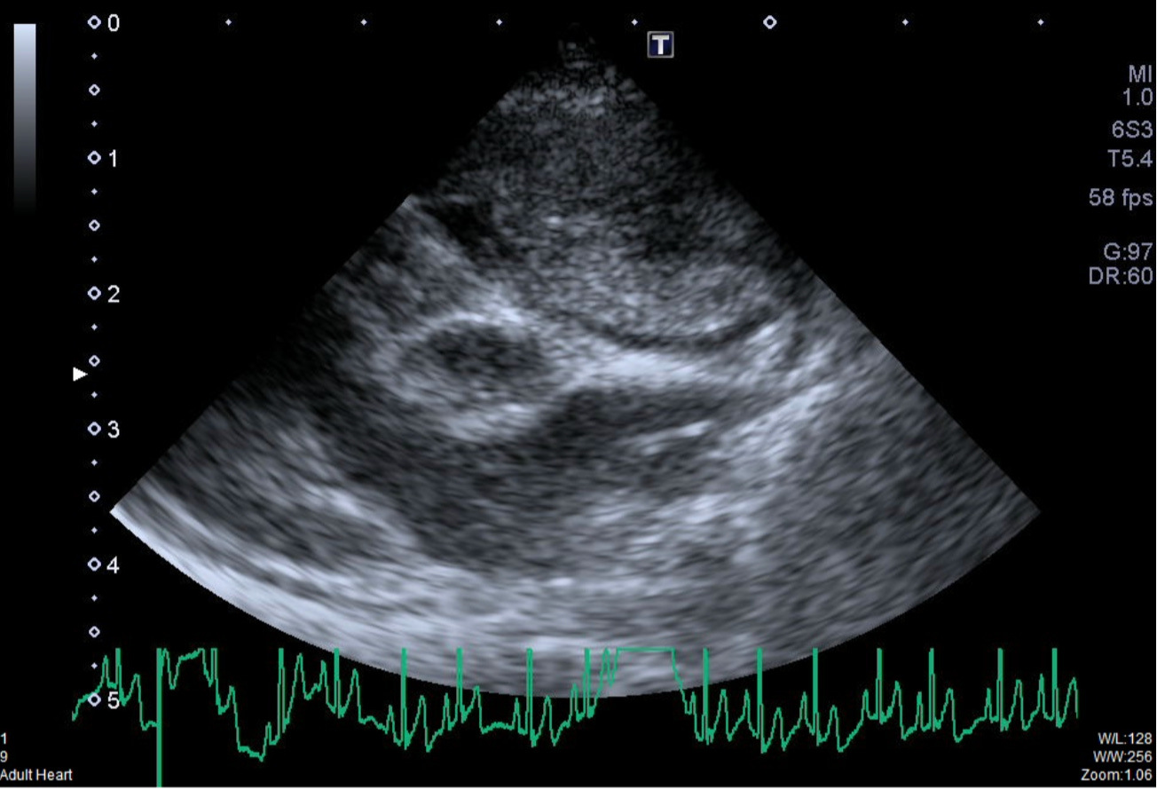
OK, so you have a patient with pericardial effusion, and you’ve stabilized them in the short term with pericardiocentesis. Great job! Now what do you do? This article will walk you through how to process and evaluate the fluid you’ve removed, and what it may (or may not) tell you.
Preliminary Analysis
The first step to any fluid is to look at it grossly and note the appearance: Does it look like frank blood or serosanguinous? Thin and straw-colored? Thick and cloudy? These, in combination with other data, can point towards distinct differentials. Next, check a PCV and total solids (TS) on the fluid, and put some of the fluid in a red top tube (no anticoagulant). Wait a few minutes to see if it clots or not; iatrogenic hemorrhage from nicking a vessel during the procedure should rapidly coagulate like normal blood, while a long-standing effusion, even if grossly very bloody, should not clot.
Any fluid with a PCV >10% is considered a hemorrhagic effusion, which is the most common cause of pericardial effusion. Recent hemorrhage is more likely to have a PCV close to peripheral blood, while chronic, insidious hemorrhage will be quite a bit lower. The TS data can help classify non-hemorrhagic effusions, and we’ll get to that in a minute.
The last core component of fluid analysis is assessing the total nucleated cell count (TNCC). This can be done by a semi-quantitative estimate from a direct fluid smear or by running the effusion through a CBC analyzer. Many—but not all!—analyzers can process cavitary effusion samples; check with your manufacturer instructions and documents.
Effusion Classifications
Pericardial effusions can be broken down into several distinct groups of diseases by mechanism as follows:
Hemorrhagic
Characteristics: >10% PCV, non-clotting blood, supported by cytology
Causes: ***Neoplasia*** (most common), idiopathic (second most common, about 20% of cases), coagulopathy, trauma, left atrial rupture (very rare)
Exudate
Characteristics: TNCC >5,000/uL, TS >2.5 g/dL
Causes: Neoplasia, septic inflammation, sterile inflammation (i.e. 2° to chronic hemorrhage, neoplasia, chyle, others)
Protein-poor (“pure”) transudate
Characteristics: TNCC <5,000/uL (usually MUCH lower), TS <2.5 g/dL
Causes: Increased hydrostatic pressure, decreased plasma oncotic pressure (severe hypoproteinemia)
Protein-rich (“modified”) transudate
Characteristics: TNCC <5,000/uL (usually MUCH lower), TS >2.5 g/dL
Causes: Vasculitis, vascular/lymphatic obstruction, heart failure (rare in dogs)
Cytology
Cytology can support the classifications above and may provide further information, i.e. identifying neoplastic cells or infectious organisms. It is always recommended to make direct or straight fluid slide(s), and further concentration by centrifugation and/or cytospin techniques (including homemade versions)1 may increase cellular yield for rare findings. As stated above, pericardial effusions are often very bloody. Several cytology findings can help discriminate peracute or iatrogenic hemorrhagic from chronic hemorrhage.
Keep reading with a 7-day free trial
Subscribe to All Science Great & Small to keep reading this post and get 7 days of free access to the full post archives.