The Continuing Education Center
CE articles, case image gallery, infographics, lectures and more!
Dear Readers,
I am always trying to add new types of content that you will find fun and informative. A few weeks ago, I polled you about your level of interest in creating a case bank for medical images. The poll question got a favorable reception by people who answered, so I decided to work on building it out. I quickly realized I had a more ambitious vision for what this could become than simply a few examples of cytology lesions.
After quietly working on this for a while now, today I am excited to launch the initial version of the All Science Great & Small CE Center!
🔬CE Center
Welcome to the Continuing Education (CE) Center! Here you will find dozens of continuing education articles and lecture presentations in addition to the case bank of images of numerous tumors, infectious organisms, and more 🤓🔬
What is the CE Center? In short, it is a central place to find all types of continuing education materials I’ve created in the past and going forward in the future. There is some FREE content in there, although much of it is bonus material for paying subscribers. NOTE: This is not an EXTRA cost on top of the existing $7/month or $70/year, it is simply giving you more bang for your buck!
For the same cost as a fancy latte a month or Netflix, you will get all of my usual email articles for free subscribers, PLUS access to the entire case bank, downloadable lecture PDFs (think of these like mini e-Books), infographics, and all of the pay-walled content I have created on this platform since it launched in 2022. What’s more is this isn’t just a static resource—I will regularly update and expand the CE center over time! Here are some details about the different sections…
Case Gallery
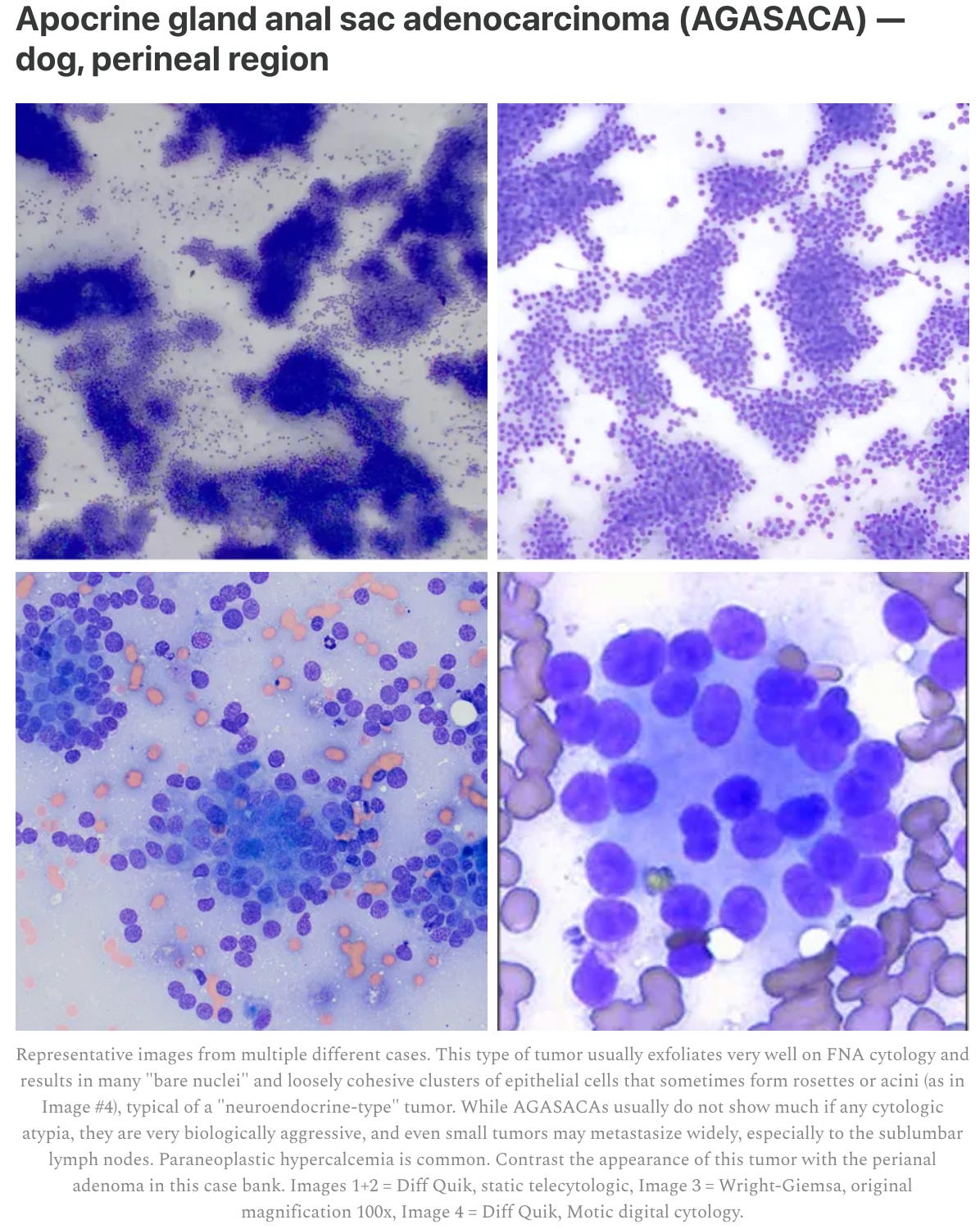
What does this look like? There are currently more than SEVENTY photomicrographs from 17 different lesion types (most include examples from multiple patients). Each lesion includes a gallery of images that can be expanded into a larger size and is accompanied by a detailed legend explaining relevant clinical and microscopic details. A big point of differentiation for this case bank is that most cases include examples in several different stains, such as Diff-Quik vs Wright-Giemsa. Furthermore, some include these images were taken on a high-resolution integrated microscope camera, while others show examples of their appearance on smart phone cameras and digital slide scanner versions, each of which has a slightly different look. The upshot is you can familiarize yourself with the many faces of specific tumors and bugs, rather than trying to squint at a single small image in a textbook that doesn’t look like what you’ll see in the real world. Finally, some of the cases include ancillary special stains, such as alkaline phosphatase for osteosarcoma and acid-fast for mycobacteria.
Here’s an example (this is just a screenshot, so you can’t expand the thumbnails):
Initial cases:
12 types of cancer
Apocrine gland anal sac adenocarcinoma (AGASACA)
Ceruminous gland adenocarcinoma
Fibrosarcoma (keloidal)
Hemangiosarcoma
Insulinoma
Liposarcoma
Lymphoma (granular/LGL) → dog spleen & cat intestinal mass
Osteosarcoma
Perianal/hepatoid gland tumor
Seminoma
Transmissible Venereal Tumor (TVT)
Transitional/urothelial carcinoma
5 types of infectious organisms
Blastomyces dermatitidis (Blastomycosis)
Cryptococcus spp. (Cryptococcosis)
Histoplasma capsulatum (Histoplasmosis)
Mycobacteria
Pythium
Continuing education lectures
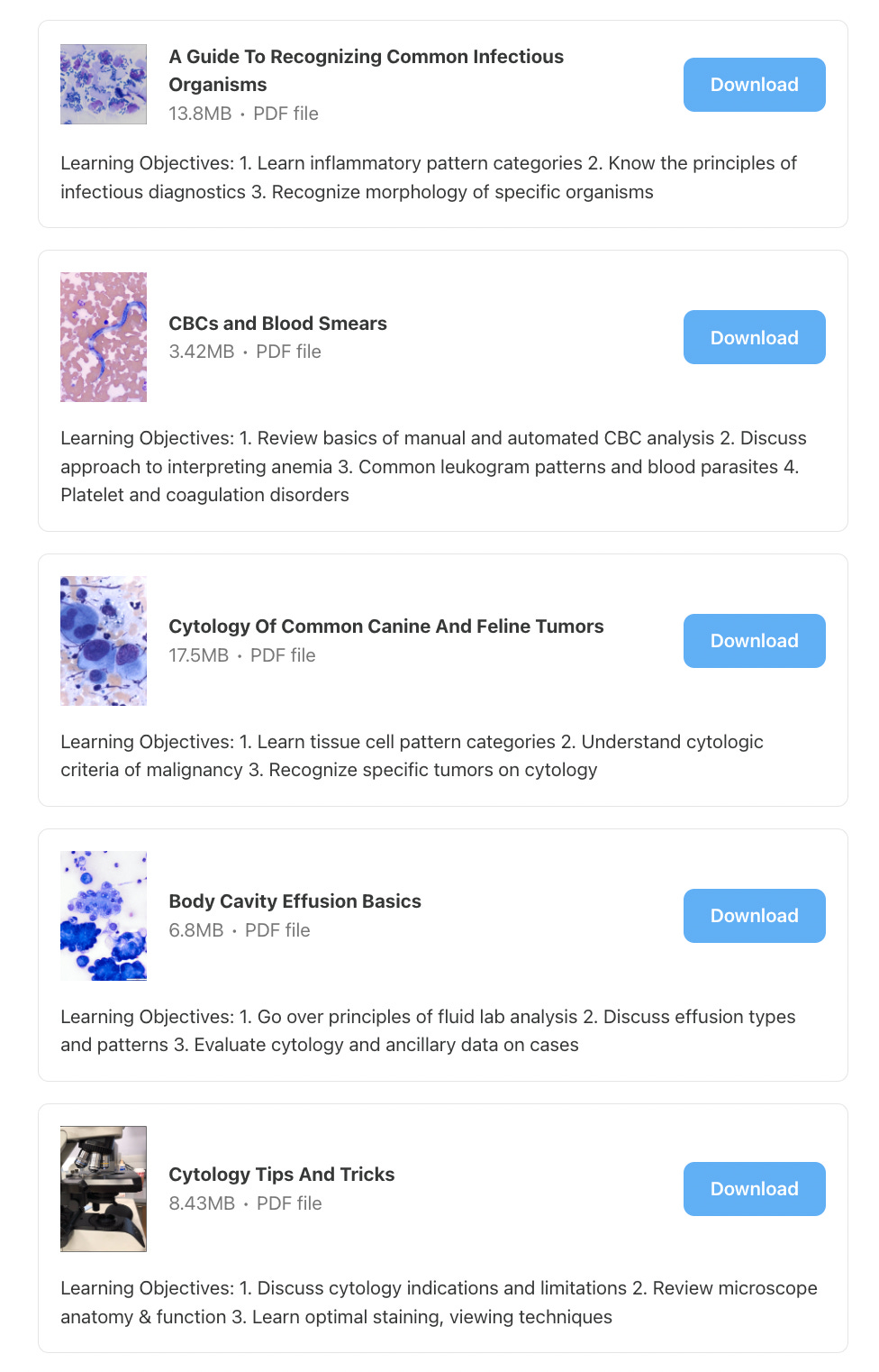
I started by uploading FIVE-HOURS worth of lectures I have previously given around the world in downloadable PDF format:
Deep-Dives on Specific Topics
Infographics
While in-depth articles are a great tool for learning, sometimes you don’t have the time to read those. And even if you do, it can be hard to remember the main points. That’s why I created these handy charts and diagrams that you can use for quick reference.
How to Choose PARR vs Flow Cytometry for Lymphoma
How to Differentiate Common Fungal Organisms
How to Prepare A Cytology Slide
Where To Look on A Cytology Slide or Blood Smear
Example:
Navigation and Future
So, how can you get to the CE Center? Besides clicking the article at the top of this post, you can find a permanent link on the main website navigation bar 👇
I want to stress that this is just the beginning for the CE Center. I considered waiting longer until I had it even more built out, but I wanted to give you access to what I had done so far. Think of this in the Silicon Valley terms of MVP, or “minimum viable product.” What do I have planned for the CE Center in the future? Lots!
For starters, I will be continually adding images and lesion types to the case gallery. There will be more tumors and infectious agents, but beyond that, I will be adding examples of normal tissues, hyperplasia, sterile inflammatory/immune-mediated lesions, examples of cysts and papillomas and more. There will be hematology images and even gross photos of lesions and laboratory findings (think urinalysis, different reactions in blood tubes, wet mount images, etc), even panels of labwork with interpretations attached. In addition to static images, I would love to include whole slide image (WSI) scans to create an experience that is essentially like reviewing an entire virtual slide without needing any equipment besides a laptop and an internet connection. (I’m currently working on solutions to some of the technical challenges for scanning and hosting those much larger files)
I also plan to add more of my infographics and continuing education lectures I’ve delivered over the years as downloadable files. Instructional videos are another type of content that I think vets, techs, and students would love.
Finally, as I continue to post deep-dive articles, I will include both free and paid types in the CE center to make them easier to find and navigate.
So there you have it! I hope you spend a few minutes checking out the great content in the CE center and that you find it worth upgrading to a paid subscription. Let me know in the comments or shoot me an email what feedback you have about the CE center so far, and any requests for new content to add!
Cheers,
—Eric


Very cool, congratulations!
In my job we have a yearly allowance/stipend to spend on (human) medical education, journals, etc. I'm not sure if this extends to the veterinary/pathology world, but if it does I would consider promoting this as a reimbursable educational/professional expenditure.
Maybe add a PDF certificate with a description of the CE Center that can be submitted to an employer?
Hard work and innovation and education should be rewarded, ideally at the same rate that professional baseball players get paid to... hit a ball.
Wow!!